Sidexis 4 Operator’s Manual PDF (Sirona Dental) DOWNLOAD
Original price was: $95.00.$32.95Current price is: $32.95.
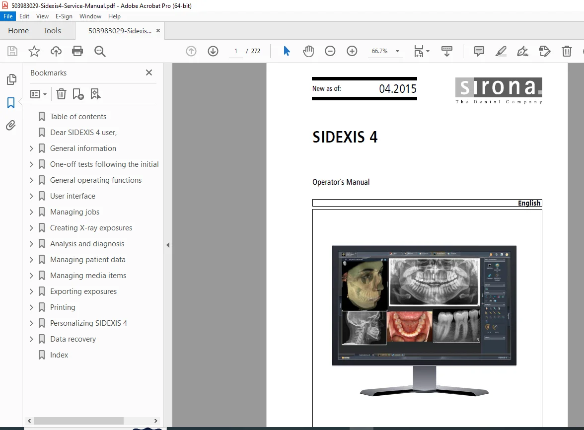
Download the Sidexis 4 operator’s manual download as a comprehensive Sirona Sidexis 4 user guide PDF for dental imaging software, covering user interface, exposure techniques, patient management, and 3D analysis. This Sidexis 4 dental software manual includes detailed instructions on measurements, personalization, and data recovery for the Sidexis 4 imaging system manual. Perfect for dental professionals seeking a PDF guide for Sidexis 4 dental X-ray software. High-quality PDF with 272 pages from 2015.
Description
Sidexis 4 Operator’s Manual PDF (Sirona Dental) DOWNLOAD
Description
The Sidexis 4 Operator’s Manual PDF is an essential Sirona Sidexis 4 user guide PDF designed for dental professionals using the Sidexis 4 dental software manual. This detailed resource provides step-by-step guidance on the user interface, managing jobs, creating X-ray exposures, analysis and diagnosis tools, patient data management, and exporting/printing functionalities within the Sidexis 4 imaging system manual. Whether you’re optimizing 3D alignments, performing measurements, or personalizing settings, this PDF guide for Sidexis 4 dental X-ray software covers safety information, system requirements, and troubleshooting to ensure seamless operation in dental practices.
Key topics include general operating functions, work phases like “Exposure” and “Examination,” image editing techniques, and Sirona dental Sidexis 4 operation instructions for intraoral exposure series, DICOM exports, and data recovery. The manual emphasizes intended use for dental diagnostics while highlighting contraindications and foreseeable misuse.
File Details:
- Manual Name: SIDEXIS 4 Operator’s Manual
- Models Covered: SIDEXIS 4 dental imaging software
- Year: 2015
- Manual PDF Quality: High-quality scanned PDF
- Number of Pages: 272
Table of Contents (Extracted and Categorized):
- Chapter 1: Introduction
- Dear SIDEXIS 4 user (Page 8)
- Chapter 2: General Information
- About this user manual (Page 9)
- General information (Page 9)
- Conventions (Page 10)
- List of abbreviations (Page 10)
- Identification of danger levels (Page 11)
- Formats and symbols used (Page 11)
- Intended use (Page 12)
- Foreseeable misuse (Page 12)
- Indication and contraindication (Page 12)
- Indications (Page 12)
- Contraindications (Page 12)
- Approval (Page 12)
- System requirements (Page 12)
- General safety information (Page 13)
- Program extensions (plug-ins) (Page 13)
- Contact information (Page 13)
- About this user manual (Page 9)
- Chapter 3: One-off Tests Following the Initial Installation
- Checking the screen output (Page 14)
- Chapter 4: General Operating Functions
- Starting / exiting the program (Page 15)
- Full-screen and window mode (Page 16)
- User manual in PDF format (Page 16)
- Chapter 5: User Interface
- Header line (Page 19)
- The phase bar (Page 20)
- “Start” work phase (Page 21)
- Job list (Page 22)
- Appointment list (Page 23)
- The “Patient” work phase (Page 24)
- The “Exposure” work phase (Page 25)
- The “Examination” work phase (Page 28)
- Work areas (Page 29)
- Tool kits (Page 33)
- The “Output” work phase (Page 45)
- “Start” work phase (Page 21)
- System menu (Page 46)
- Configuration menu (Page 47)
- “General settings” (Page 50)
- “Patient settings” (Page 55)
- “Exposure” (Page 56)
- “Output” (Page 63)
- “SIDEXIS Manager” (Page 65)
- Configuration menu (Page 47)
- Docking window (Page 66)
- Session bar (Page 69)
- General control elements (Page 70)
- Call up shortcut menus (Page 72)
- Shortcut keys (Page 73)
- General shortcut keys (Page 73)
- Shortcut keys within the timeline (Page 75)
- Chapter 6: Managing Jobs
- Creating an X-ray job (Page 78)
- Accepting X-ray jobs (Page 79)
- Accepting X-ray jobs automatically (Page 79)
- Accepting X-ray jobs manually (Page 79)
- Sorting the job list (Page 80)
- Chapter 7: Creating X-ray Exposures
- Selecting an X-ray job (Page 83)
- Selecting an X-ray device (Page 85)
- Entering an indication (Page 87)
- Taking X-ray exposures on the device (Page 89)
- Repeat the exposure (Page 90)
- Intraoral exposure series (Page 91)
- Example of performing an intraoral exposure series (Page 93)
- Stopping / starting (pausing) exposure series in the series dialog (Page 96)
- Repeating an exposure in the series dialog (Page 97)
- Canceling an exposure series (Page 99)
- Continuing an exposure series (Page 101)
- Chapter 8: Analysis and Diagnosis
- Work areas for 2D images, 3D volumes, and section views (Page 104)
- “3D examination” (Page 105)
- Examination window (Page 106)
- Position aids (Page 109)
- Standard layouts (Page 111)
- Panoramic image (Page 117)
- 3D image (Page 118)
- Section views (Page 124)
- Opening a new “3D examination” work area (Page 135)
- “Light box” (Page 136)
- Opening a new “light box” work area (Page 137)
- Working with media windows (Page 138)
- “Compare” (Page 148)
- Opening a new “compare” work area (Page 150)
- “3D examination” (Page 105)
- Opening last exposures (Page 152)
- Continue last patient session (Page 153)
- Saving / opening patient sessions (Page 154)
- Set panoramic curve (Page 158)
- Opening the “panoramic curve editor” menu (Page 159)
- Moving the panoramic curve to another section plane (Page 159)
- Automatically adjusting panoramic curves to preset dentition shapes and sizes (Page 160)
- Editing the panoramic curve manually (Page 161)
- 3D alignment (Page 164)
- Opening the “3D alignment” menu (Page 164)
- Correcting positions (Page 165)
- Editing images (Page 167)
- Brightness / contrast / tonal value (Page 167)
- Setting the brightness / contrast via the mouse controller (Page 168)
- Automatic contrast optimization (Page 169)
- Setting the brightness / contrast and tonal value through the dials (Page 169)
- Resetting the settings for brightness, contrast, and tonal value (Page 170)
- Image filters (Page 171)
- Relief (Page 171)
- Applying sharpness (Page 172)
- Reducing noise (Page 172)
- Smooth down (Page 173)
- Resetting / hiding an image filter (Page 174)
- Rotating (Page 176)
- Brightness / contrast / tonal value (Page 167)
- Performing measurements (Page 177)
- Angles (Page 177)
- Measuring angles (Page 177)
- Editing angle sizes (Page 180)
- Lengths (Page 181)
- Measuring lengths (Page 181)
- Editing paths (Page 183)
- Reference measurement (Page 184)
- Measuring a reference object (Page 184)
- Editing reference measurements (Page 186)
- Angles (Page 177)
- Creating and editing remarks (Page 187)
- Creating remarks (Page 188)
- Retrieving notes in section views (Page 189)
- Moving remarks (Page 190)
- Moving dimension indicators (Page 191)
- Changing the color of the remarks (Page 192)
- Showing/hiding remarks (Page 193)
- Delete notes (Page 193)
- Optimizing the 3D image display (Page 194)
- Setting the bone threshold value (Page 194)
- Setting surface transparency for FaceScan exposures (Page 196)
- Transfer function editor (Page 198)
- Documenting findings (Page 199)
- Creating a new diagnosis (Page 200)
- Work areas for 2D images, 3D volumes, and section views (Page 104)
- Chapter 9: Managing Patient Data
- Working with the patient table (Page 204)
- Displaying patients according to consultant (Page 205)
- Sorting a patient table (Page 205)
- Free text search in the patient table (Page 206)
- Create a new patient (Page 207)
- Displaying patient details (Page 209)
- Editing patient details (Page 210)
- Logging patients in/out (Page 212)
- Working with the patient table (Page 204)
- Chapter 10: Managing Media Items
- Timeline and media gallery (Page 213)
- Filtering exposures according to region (Page 215)
- Moving along the timeline (Page 217)
- Opening exposures for examination (Page 218)
- Opening individual media items (Page 218)
- Opening multiple media items in the work area simultaneously (Page 219)
- Importing media (Page 222)
- Importing media items manually (Page 223)
- Automatic import via directory monitoring (Page 226)
- Importing duplicates (Page 229)
- Exposures from TWAIN data sources (Page 230)
- Important information on scanning X-ray images (Page 231)
- Deleting media items (Page 232)
- Reactivating deleted media items (Page 233)
- Chapter 11: Exporting Exposures
- Exporting 2D views (Page 235)
- Sending 2D views by email (Page 238)
- DICOM Export Wrap&Go (Page 240)
- Exporting DICOM examination export and volumes (Page 242)
- Chapter 12: Printing
- Printing an image (Page 245)
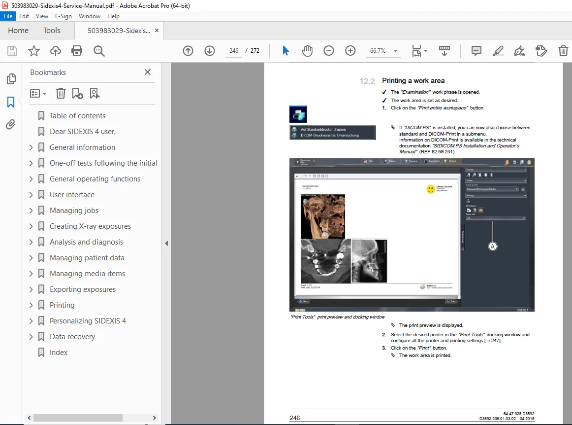
- Printing a work area (Page 246)
- The “Printing tools” docking window (Page 247)
- Chapter 13: Personalizing SIDEXIS 4
- Call up the configuration menu (Page 248)
- Setting the program language (Page 249)
- Defining the practice address and practice logo for printouts (Page 251)
- Selecting the dental notation (Page 256)
- User set up/administration (Page 258)
- Configuring patient display (Page 260)
- Chapter 14: Data Recovery
- Rescue program (Sirona Control Admin Rescue) (Page 262)
- Starting the “Sirona Control Admin Rescue” program (Page 263)
- Requesting data (Page 265)
- Rescue program (Sirona Control Admin Rescue) (Page 262)
- Index (Page 266)
This PDF guide for Sidexis 4 dental X-ray software equips you with Sirona dental Sidexis 4 operation instructions to enhance diagnostic accuracy and workflow efficiency.
Unlock efficient dental imaging today—download this Sidexis 4 operator’s manual download instantly and master your Sirona system with ease!