★ TRUSTED BY 321,897+ PROFESSIONALS WORLDWIDE
🏢
Trusted Business
Verified & Licensed
🛡️
Virus Free Files
100% Safe Downloads
🔒
Secure Payment
SSL Protected
Instant Delivery
Available Immediately
Sale!

Medtronic Advisa DR MRI SureScan A3DR01 & Advisa SR MRI A3SR01 Clinician Manual PDF

Original price was: $24.95.Current price is: $14.95.

Full 352-page Clinician Manual (December 2017) for the Medtronic Advisa DR MRI SureScan A3DR01 dual chamber and Advisa SR MRI SureScan A3SR01 single chamber pacemakers, covering programming, implant, follow-up, and quick-reference specification data. Instant PDF download.

Instant PDF Download
Available immediately
💾
Save to Your Device
Download & keep forever
🛡️
Antivirus Scanned
100% virus-free
🌍
Trusted Worldwide
175,000+ customers

Description

Medtronic Advisa DR MRI SureScan A3DR01 & Advisa SR MRI A3SR01 Clinician Manual

This is the complete Clinician Manual for the Medtronic Advisa DR MRI SureScan A3DR01 dual chamber pacemaker and the Advisa SR MRI SureScan A3SR01 single chamber pacemaker, dated December 2017. The document runs 352 pages and is the primary operating and programming reference used with the Medtronic programmer for both device models.

  • Manual type: Clinician Manual
  • Brand / Models: Medtronic Advisa DR MRI SureScan A3DR01 (dual chamber, DDDR) and Advisa SR MRI SureScan A3SR01 (single chamber, VVIR)
  • Language: English
  • Page count: 352 pages
  • File format: PDF
  • MAPS ID: 502714-019
  • Document date: December 2017

Chapters Covered

  • System overview — introduction, Advisa DR and SR system description, feature model matrix, indications and usage, contraindications, MRI conditions for use
  • Warnings, precautions, and potential adverse events — general warnings, explant and disposal, handling and storage, lead evaluation and connection, device operation
  • Using the programmer — establishing telemetry, conducting a patient session, display screen features, emergency VVI pacing, Checklist workflow, viewing and programming parameters, TherapyGuide, patient information, Live Rhythm Monitor, saving device data, printing reports
  • Advisa DR MRI implant procedures — preparing for implant, selecting and implanting leads, testing the lead system, connecting leads to the device, positioning and securing the device, replacing a device
  • Advisa SR MRI implant procedures — the same implant workflow for the single chamber device
  • Conducting a patient follow-up session — follow-up guidelines, stored data summary, Cardiac Compass Report, heart failure management, OptiVol fluid monitoring, Arrhythmia Episodes data, episode and therapy counters, Flashback Memory, Rate Drop Response episodes, rate histograms, lead performance data, automatic device status monitoring, optimizing device longevity
  • Configuring pacing therapies — sensing intrinsic activity, pacing therapies, MVP mode, rate-responsive pacing, Capture Management, lead polarity, AV interval adaptation, PVARP adjustment, Rate Drop Response, PVC Response, Ventricular Rate Stabilization, atrial intervention pacing
  • Configuring tachyarrhythmia detection and therapies — atrial and ventricular tachyarrhythmia detection, suspending/resuming detection, scheduling atrial therapies, antitachycardia pacing
  • Testing the system — evaluating underlying rhythm, measuring pacing thresholds and lead impedance, sensing test, magnet mode operation, inducing an arrhythmia, delivering a manual therapy
  • Appendix A/B — Advisa DR and SR quick reference: physical characteristics, electrical specifications, replacement indicators, projected service life, magnet application, stored data and diagnostics
  • Appendix C/D — Advisa DR and SR device parameters: emergency settings, tachyarrhythmia detection parameters, therapy parameters, pacing parameters, data collection parameters, system test parameters, EP Study parameters, nonprogrammable parameters
  • Glossary and Index

IMAGES PREVIEW

Medtronic Advisa DR MRI SureScan A3DR01 & Advisa SR MRI A3SR01 Clinician Manual PDF — PDF page 1 preview
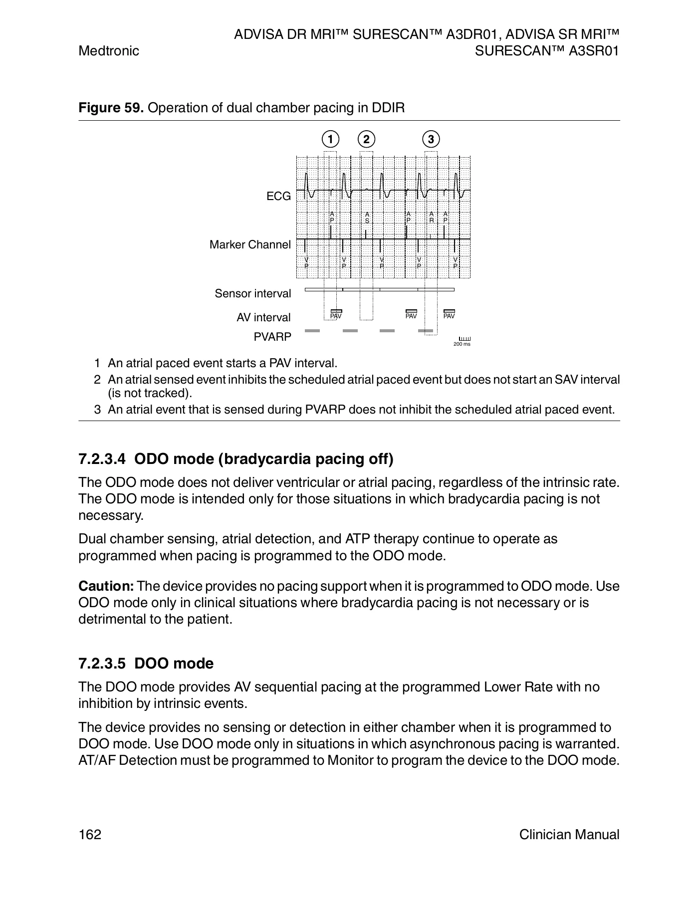
Medtronic Advisa DR MRI SureScan A3DR01 & Advisa SR MRI A3SR01 Clinician Manual PDF — PDF page 162 preview
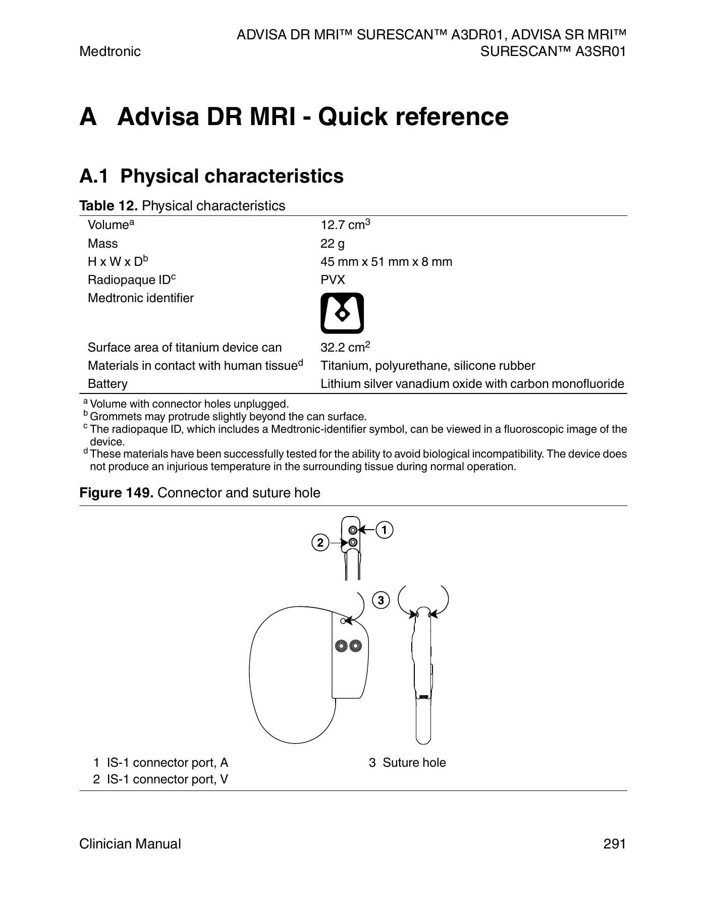
Medtronic Advisa DR MRI SureScan A3DR01 & Advisa SR MRI A3SR01 Clinician Manual PDF — PDF page 291 preview

Taken together, this Clinician Manual gives the reader a complete working reference for programming and managing both Advisa MRI SureScan device models — from initial telemetry setup and implant technique through long-term follow-up, tachyarrhythmia management, and end-of-life replacement planning.

Complete PDF Bookmark Tree

Complete Bookmarks

The complete bookmarks in the “Medtronic Advisa DR MRI SureScan A3DR01 & Advisa SR MRI A3SR01 Clinician Manual PDF” are as follows:

Contentsp. 5
1 System overviewp. 10
1.1 Introductionp. 10
1.1.1 CE mark of conformityp. 10
1.1.2 About this manualp. 10
1.1.2.1 Manual conventionsp. 11
1.1.3 Product literaturep. 11
1.1.4 Technical supportp. 12
1.1.5 Customer educationp. 13
1.1.6 Noticep. 13
1.2 Advisa DR MRI system descriptionp. 13
1.3 Advisa SR MRI system descriptionp. 15
1.4 Advisa DR and SR MRI feature model matrixp. 17
1.5 Indications and usagep. 18
1.6 Contraindicationsp. 19
1.7 MRI conditions for usep. 19
2 Warnings, precautions, and potential adverse eventsp. 21
2.1 General warnings and precautionsp. 21
2.2 Explant and disposalp. 22
2.3 Handling and storage instructionsp. 22
2.3.1 Device handlingp. 22
2.3.2 Device storagep. 23
2.4 Lead evaluation and lead connectionp. 23
2.5 Device operationp. 23
2.5.1 Pacemaker-dependent patientsp. 26
2.6 Potential adverse eventsp. 26
3 Using the programmerp. 28
3.1 Establishing telemetry between the device and the programmerp. 28
3.1.1 How to establish telemetry between the device and the programmerp. 28
3.1.2 How to maintain reliable telemetryp. 28
3.2 Conducting a patient sessionp. 29
3.2.1 Starting a patient sessionp. 29
3.2.1.1 How to start a patient sessionp. 29
3.2.2 Device and telemetry effects during a patient sessionp. 29
3.2.3 How to interrogate the device during the sessionp. 30
3.2.4 Ending a patient sessionp. 30
3.2.4.1 How to end a patient sessionp. 30
3.3 Display screen featuresp. 31
3.3.1 Task barp. 32
3.3.2 Status barp. 32
3.3.3 Live Rhythm Monitor windowp. 33
3.3.4 Task areap. 33
3.3.5 Tool palettep. 34
3.3.6 Buttonsp. 35
3.3.7 Command barp. 35
3.4 Enabling emergency VVI pacingp. 36
3.4.1 Considerations for emergency VVI pacingp. 36
3.4.2 How to enable emergency VVI pacingp. 36
3.5 Streamlining implant and follow-up sessions with Checklistp. 37
3.5.1 How to use a standard checklistp. 37
3.5.2 How to create and edit a custom checklistp. 38
3.6 Viewing and programming device parametersp. 39
3.6.1 Understanding the symbols used on the Parameters screenp. 39
3.6.2 How to access parametersp. 41
3.6.3 How to access a group of related parametersp. 42
3.7 Saving and retrieving a set of parameter valuesp. 43
3.7.1 How to save a set of parameter valuesp. 43
3.7.2 How to retrieve a set of parameter valuesp. 44
3.8 Using TherapyGuide to select parameter valuesp. 44
3.8.1 Operation of TherapyGuidep. 45
3.8.2 Considerations for TherapyGuidep. 46
3.8.3 How to obtain a set of suggested valuesp. 47
3.8.4 How to view the rationale for TherapyGuide suggestionsp. 48
3.9 Viewing and entering patient informationp. 48
3.9.1 How to view and enter patient informationp. 49
3.9.2 How to export saved lead measurements to the Implant windowp. 51
3.10 Working with the Live Rhythm Monitorp. 52
3.10.1 Viewing live waveform tracesp. 53
3.10.1.1 How to select and adjust the waveformsp. 53
3.10.1.2 How to change the appearance of the waveformp. 54
3.10.1.3 How to interpret Marker Channel annotations and symbolsp. 55
3.10.2 Recording live waveform tracesp. 56
3.10.3 Freezing live waveform tracesp. 57
3.10.4 Recalling waveform stripsp. 58
3.10.4.1 How to recall a waveform stripp. 59
3.11 Saving and retrieving device datap. 59
3.11.1 Saving device datap. 60
3.11.1.1 Considerations for saving device data on a USB flash drivep. 60
3.11.1.2 How to save device data to a USB flash drivep. 61
3.11.1.3 Preparing to save data to a diskp. 61
3.11.1.4 How to save device data to a diskp. 61
3.11.2 Retrieving device datap. 61
3.11.2.1 How to read device data from a USB flash drive or a diskp. 62
3.12 Printing reportsp. 62
3.12.1 Setting preferences for printing, reports, and testsp. 62
3.12.1.1 How to set printing preferencesp. 63
3.12.2 Printing an Initial Interrogation Reportp. 63
3.12.2.1 How to set Initial Report preferencesp. 64
3.12.3 Printing reports during a patient sessionp. 64
3.12.3.1 How to print a customized set of reportsp. 65
3.12.3.2 How to print a report on a specific programming screenp. 65
3.12.4 Printing a summary report for the patient sessionp. 66
3.12.4.1 How to print a summary report for the patient sessionp. 66
3.12.4.2 How to set Final Report preferencesp. 66
3.12.5 Managing the Print Queuep. 67
3.12.5.1 How to use the Print Queue window during a patient sessionp. 67
3.12.5.2 How to use the Print Queue window outside of a patient sessionp. 67
3.12.5.3 Interpreting the Print Queue Status columnp. 68
3.12.6 Setting Tests preferencesp. 68
3.12.6.1 How to set Tests preferencesp. 69
4 Advisa DR MRI – Implanting the devicep. 70
4.1 Preparing for an implantp. 70
4.1.1 Instruments, components, and accessories required for an implantp. 70
4.1.2 Setting up the programmer and starting the applicationp. 70
4.1.3 Considerations for preparing for an implantp. 71
4.1.4 How to prepare the device for implantp. 72
4.2 Selecting and implanting the leadsp. 73
4.2.1 Selecting the leadsp. 73
4.2.2 How to verify lead and connector compatibilityp. 73
4.2.3 Implanting the leadsp. 74
4.3 Testing the lead systemp. 74
4.3.1 Considerations for testing the lead systemp. 74
4.3.2 How to verify and save the sensing and pacing valuesp. 74
4.4 Connecting the leads to the devicep. 76
4.4.1 How to connect a lead to the devicep. 76
4.5 Positioning and securing the devicep. 78
4.5.1 How to position and secure the devicep. 78
4.6 Completing the implant procedurep. 79
4.6.1 How to complete programming the devicep. 79
4.6.2 How to assess the performance of the device and leadsp. 80
4.7 Replacing a devicep. 80
4.7.1 How to explant and replace a devicep. 81
5 Advisa SR MRI – Implanting the devicep. 82
5.1 Preparing for an implantp. 82
5.1.1 Instruments, components, and accessories required for an implantp. 82
5.1.2 Setting up the programmer and starting the applicationp. 83
5.1.3 Considerations for preparing for an implantp. 83
5.1.4 How to prepare the device for implantp. 84
5.2 Selecting and implanting the leadsp. 85
5.2.1 Selecting the leadp. 85
5.2.2 How to verify lead and connector compatibilityp. 85
5.2.3 Implanting the leadp. 85
5.3 Testing the lead systemp. 86
5.3.1 Considerations for testing the lead systemp. 86
5.3.2 How to verify and save the sensing and pacing valuesp. 86
5.4 Connecting the lead to the devicep. 87
5.4.1 How to connect a lead to the devicep. 88
5.5 Positioning and securing the devicep. 89
5.5.1 How to position and secure the devicep. 89
5.6 Completing the implant procedurep. 91
5.6.1 How to complete programming the devicep. 91
5.6.2 How to assess the performance of the device and leadp. 91
5.7 Replacing a devicep. 91
5.7.1 How to explant and replace a devicep. 92
6 Conducting a patient follow-up sessionp. 94
6.1 Patient follow-up guidelinesp. 94
6.1.1 Follow-up toolsp. 94
6.1.2 Reviewing the presenting rhythmp. 95
6.1.3 Verifying the status of the implanted systemp. 95
6.1.3.1 How to review the battery voltage and device status indicatorsp. 95
6.1.3.2 How to assess the performance of the device and leadsp. 95
6.1.4 Verifying the clinical effectiveness of the implanted systemp. 96
6.1.4.1 How to assess effective pacing therapyp. 96
6.1.4.2 How to assess accurate tachyarrhythmia detectionp. 96
6.1.4.3 How to assess appropriate tachyarrhythmia therapyp. 97
6.2 Viewing a summary of recently stored datap. 97
6.2.1 How to view the Quick Look II screenp. 97
6.2.2 Information provided by the Quick Look II screenp. 98
6.2.2.1 Assessing the device and lead statusp. 98
6.2.2.2 Assessing the patient’s conditionp. 99
6.2.2.3 Quick Look II Observationsp. 100
6.3 Viewing long-term clinical trends with the Cardiac Compass Reportp. 101
6.3.1 How to view and print Cardiac Compass Trendsp. 101
6.3.2 Information provided by the Cardiac Compass Reportp. 102
6.3.2.1 Event informationp. 102
6.3.2.2 Assessing AT/AF arrhythmia informationp. 103
6.3.2.3 Assessing pacing and rate response informationp. 104
6.3.2.4 Assessing heart failure informationp. 105
6.4 Viewing heart failure management informationp. 106
6.4.1 How to print the Heart Failure Management Reportp. 106
6.4.2 Information provided by the Heart Failure Management Reportp. 107
6.4.2.1 Patient informationp. 107
6.4.2.2 Clinical Status and Observationsp. 107
6.4.2.3 Event informationp. 108
6.4.2.4 Assessing OptiVol fluid trendsp. 108
6.4.2.5 Assessing clinical trendsp. 109
6.5 Monitoring for thoracic fluid accumulation with OptiVolp. 111
6.5.1 System solution: OptiVol Fluid Status Monitoringp. 112
6.5.2 Operation of OptiVol Fluid Status Monitoringp. 112
6.5.2.1 Daily and Reference Impedancesp. 112
6.5.2.2 SentryCheck Monitorp. 114
6.5.3 Programming considerations for OptiVol Fluid Status Monitoringp. 114
6.5.4 Programming OptiVol Fluid Status Monitoringp. 115
6.5.4.1 Programming the OptiVol Thresholdp. 115
6.5.5 Evaluation of OptiVol Fluid Status Monitoringp. 115
6.5.5.1 Viewing OptiVol fluid trendsp. 115
6.5.5.2 Viewing OptiVol observationsp. 116
6.5.5.3 Viewing the OptiVol event logp. 117
6.6 Viewing Arrhythmia Episodes data and setting data collection preferencesp. 117
6.6.1 How to view the Arrhythmia Episodes datap. 117
6.6.2 Viewing the episode logp. 117
6.6.3 Viewing episode recordsp. 119
6.6.3.1 Viewing the episode interval plotp. 120
6.6.3.2 Viewing the episode EGMp. 121
6.6.3.3 Viewing the episode textp. 122
6.6.4 How to set data collection preferencesp. 123
6.6.4.1 Programming data collection preferencesp. 124
6.7 Viewing episode and therapy countersp. 124
6.7.1 How to view the countersp. 125
6.7.2 VT/VF episode countersp. 125
6.7.3 AT/AF episode countersp. 126
6.7.4 AT/AF therapy countersp. 127
6.8 Viewing Flashback Memory datap. 128
6.8.1 How to view Flashback Memory datap. 128
6.9 Viewing Rate Drop Response episodesp. 129
6.9.1 How to view Rate Drop Response episode datap. 130
6.10 Using rate histograms to assess heart ratesp. 131
6.10.1 How to view and print Rate Histogramsp. 131
6.10.2 Information provided by the Rate Histograms Reportp. 132
6.11 Viewing detailed device and lead performance datap. 134
6.11.1 Viewing battery and lead measurement datap. 134
6.11.1.1 How to view battery and lead measurement datap. 134
6.11.1.2 Battery voltage and replacement indicatorsp. 135
6.11.1.3 Remaining longevity estimatesp. 136
6.11.1.4 Sensing Integrity Counterp. 137
6.11.1.5 Atrial Lead Position Check resultsp. 137
6.11.1.6 Lead impedance and sensing amplitude measurementsp. 137
6.11.2 Viewing lead impedance trendsp. 137
6.11.2.1 How to view lead impedance trendsp. 138
6.11.3 Viewing sensing amplitude trendsp. 139
6.11.3.1 How to view sensing amplitude trendsp. 139
6.11.4 Viewing capture threshold trendsp. 140
6.11.4.1 How to view capture threshold trendsp. 140
6.12 Automatic device status monitoringp. 142
6.12.1 Definitions of device status indicator warningsp. 142
6.12.2 How to respond to the device status indicator warning for electrical resetp. 143
6.13 Optimizing device longevityp. 144
6.13.1 Promoting intrinsic AV conductionp. 145
6.13.2 Managing pacing outputsp. 145
6.13.3 Considering how diagnostic features with data storage impact longevityp. 146
7 Configuring pacing therapiesp. 147
7.1 Sensing intrinsic cardiac activityp. 147
7.1.1 System solution: sensingp. 147
7.1.2 Operation of sensing thresholdsp. 148
7.1.2.1 Bipolar sensingp. 148
7.1.2.2 Unipolar sensingp. 149
7.1.3 Operation of blanking periodsp. 149
7.1.4 Operation of Post-Ventricular Atrial Blanking (PVAB)p. 151
7.1.4.1 PVAB operation with bipolar atrial sensingp. 151
7.1.4.2 PVAB operation with unipolar atrial sensingp. 152
7.1.5 Operation of refractory periodsp. 153
7.1.5.1 Post Ventricular Atrial Refractory Period (PVARP)p. 154
7.1.5.2 Atrial Refractory Periodp. 154
7.1.6 Programming considerations for sensingp. 154
7.1.7 Programming sensingp. 157
7.1.7.1 Programming sensitivities, polarities, and blanking periodsp. 157
7.1.7.2 Programming refractory periodsp. 157
7.1.8 Evaluation of sensingp. 157
7.1.8.1 Using the Sensing Test to evaluate sensingp. 157
7.1.8.2 Viewing the Sensing Integrity Counterp. 158
7.1.8.3 Viewing P-wave and R-wave amplitude trendsp. 158
7.2 Providing pacing therapiesp. 159
7.2.1 System solution: pacing therapiesp. 159
7.2.2 Operation of pacing and sensingp. 160
7.2.3 Operation of dual chamber pacingp. 160
7.2.3.1 AAIRDDDR and AAIDDD modesp. 160
7.2.3.2 DDDR and DDD modesp. 160
7.2.3.3 DDIR and DDI modesp. 161
7.2.3.4 ODO mode (bradycardia pacing off)p. 162
7.2.3.5 DOO modep. 162
7.2.4 Operation of single chamber pacingp. 163
7.2.4.1 AAIRDDDR and AAIDDD modesp. 163
7.2.4.2 VVIR and VVI modesp. 163
7.2.4.3 AAIR and AAI modesp. 163
7.2.4.4 VOO modep. 164
7.2.4.5 OVO mode (bradycardia pacing off)p. 164
7.2.4.6 AOO modep. 165
7.2.5 Programming considerations for pacing therapiesp. 165
7.2.5.1 Pacing mode selectionp. 165
7.2.5.2 Programming considerations for dual chamber pacingp. 165
7.2.6 Programming pacing therapiesp. 166
7.2.7 Evaluation of pacing therapiesp. 166
7.2.8 Tracking rapid atrial ratesp. 167
7.2.8.1 2:1 blockp. 167
7.2.8.2 Upper Tracking Ratep. 168
7.3 Reducing unnecessary ventricular pacing with MVP modep. 169
7.3.1 System solution: MVP modep. 170
7.3.2 Operation of MVP modep. 170
7.3.2.1 Intact AV conductionp. 170
7.3.2.2 Loss of AV conductionp. 171
7.3.2.3 AV conduction resumesp. 171
7.3.2.4 Complete AV blockp. 172
7.3.2.5 Transient loss of conductionp. 173
7.3.2.6 Interactions with MVP modep. 173
7.3.3 Programming considerations for MVP modep. 174
7.3.4 Programming MVP modep. 174
7.3.5 Evaluation of MVP modep. 174
7.3.5.1 Status barp. 174
7.3.5.2 Quick Look II screenp. 175
7.3.5.3 Cardiac Compass Reportp. 175
7.3.5.4 Rate Histograms Reportp. 176
7.4 Providing rate-responsive pacingp. 176
7.4.1 System solution: Rate Responsep. 176
7.4.2 Operation of Rate Responsep. 176
7.4.2.1 Activity sensingp. 177
7.4.2.2 Rate calculationp. 177
7.4.2.3 Rate Profile Optimizationp. 178
7.4.2.4 Activity Acceleration and Activity Decelerationp. 179
7.4.2.5 Rate Response during implantp. 179
7.4.2.6 Rate Response parameters screenp. 180
7.4.3 Programming considerations for Rate Responsep. 180
7.4.4 Programming Rate Responsep. 181
7.4.5 Evaluation of Rate Responsep. 181
7.4.5.1 Rate Histograms Reportp. 181
7.4.5.2 Flashback Memoryp. 182
7.5 Managing pacing output energies with Capture Managementp. 183
7.5.1 System solution: Capture Managementp. 183
7.5.2 Operation of Capture Managementp. 184
7.5.2.1 Manual adjustment of pacing outputsp. 184
7.5.2.2 Pacing thresholds and safety marginsp. 184
7.5.3 Operation of Atrial Capture Managementp. 185
7.5.3.1 Preparing for an atrial pacing threshold searchp. 186
7.5.3.2 Searching for and determining the atrial pacing thresholdp. 186
7.5.3.3 Adjusting atrial pacing outputsp. 188
7.5.3.4 Stopping an atrial pacing threshold search in progressp. 189
7.5.4 Operation of Right Ventricular Capture Managementp. 189
7.5.4.1 Preparing for an RV pacing threshold searchp. 190
7.5.4.2 Searching for and determining the RV pacing thresholdp. 190
7.5.4.3 Adjusting the RV pacing outputsp. 191
7.5.4.4 Stopping an RV pacing threshold search in progressp. 192
7.5.5 Programming considerations for Capture Managementp. 192
7.5.6 Programming Capture Managementp. 193
7.5.7 Evaluation of Capture Managementp. 194
7.5.7.1 Quick Look IIp. 194
7.5.7.2 Capture Threshold trendsp. 194
7.6 Configuring lead polarityp. 195
7.6.1 System solution: Automatic Polarity Configuration and Lead Monitorp. 195
7.6.2 Operation of Automatic Polarity Configurationp. 196
7.6.3 Operation of Lead Monitorp. 196
7.6.4 Programming considerations for lead polarityp. 197
7.6.5 Programming lead polarityp. 198
7.6.6 Evaluation of lead polarityp. 198
7.7 Adapting the AV interval during rate changesp. 199
7.7.1 System solution: Rate Adaptive AVp. 199
7.7.2 Operation of Rate Adaptive AVp. 199
7.7.3 Programming considerations for Rate Adaptive AVp. 200
7.7.4 Programming Rate Adaptive AVp. 201
7.8 Adjusting PVARP to changes in the patient’s heart ratep. 201
7.8.1 System solution: Auto PVARPp. 201
7.8.2 Operation of Auto PVARPp. 201
7.8.3 Programming considerations for Auto PVARPp. 203
7.8.4 Programming Auto PVARPp. 204
7.9 Treating syncope with Rate Drop Responsep. 204
7.9.1 System solution: Rate Drop Responsep. 204
7.9.2 Operation of Rate Drop Responsep. 204
7.9.2.1 Detectionp. 205
7.9.2.2 Intervention and step-downp. 207
7.9.3 Programming considerations for Rate Drop Responsep. 208
7.9.4 Programming Rate Drop Responsep. 208
7.9.5 Evaluation of Rate Drop Responsep. 209
7.10 Promoting the intrinsic rate during periods of inactivityp. 210
7.10.1 System solution: Rate Hysteresisp. 210
7.10.2 Operation of Rate Hysteresisp. 210
7.10.3 Programming considerations for Rate Hysteresisp. 211
7.10.4 Programming Rate Hysteresisp. 211
7.10.5 Evaluation of Rate Hysteresisp. 212
7.10.5.1 Viewing the Ventricular Rate Histogram Reportp. 212
7.11 Providing a slower pacing rate during periods of sleepp. 212
7.11.1 System solution: Sleep featurep. 212
7.11.2 Operation of the Sleep featurep. 212
7.11.3 Programming considerations for the Sleep featurep. 213
7.11.3.1 How to set the device clockp. 213
7.11.4 Programming the Sleep featurep. 214
7.11.5 Evaluation of the Sleep featurep. 214
7.11.5.1 Viewing or printing Ventricular Rate Histogramsp. 214
7.11.5.2 Viewing or printing the Cardiac Compass Trends Reportp. 214
7.12 Preventing competitive atrial pacingp. 214
7.12.1 System solution: NCAPp. 215
7.12.2 Operation of NCAPp. 215
7.12.3 Programming NCAPp. 216
7.12.4 Evaluation of NCAPp. 216
7.13 Interrupting pacemaker-mediated tachycardiasp. 216
7.13.1 System solution: PMT Interventionp. 216
7.13.2 Operation of PMT Interventionp. 216
7.13.3 Programming PMT Interventionp. 217
7.14 Managing retrograde conduction using PVC Responsep. 217
7.14.1 System solution: PVC Responsep. 218
7.14.2 Operation of PVC Responsep. 218
7.14.3 Programming PVC Responsep. 219
7.15 Reducing inappropriate ventricular inhibition using VSPp. 219
7.15.1 System solution: VSPp. 219
7.15.2 Operation of VSPp. 219
7.15.3 Programming considerations for VSPp. 220
7.15.4 Programming VSPp. 220
7.15.5 Evaluation of VSPp. 221
7.16 Preventing rapid ventricular pacing during atrial tachyarrhythmiasp. 221
7.16.1 System solution: Mode Switchp. 221
7.16.2 Operation of Mode Switchp. 222
7.16.2.1 Interactions with other device operationsp. 223
7.16.3 Programming considerations for Mode Switchp. 223
7.16.4 Programming Mode Switchp. 224
7.16.4.1 Programming the Atrial Interval (Rate)p. 224
7.16.5 Evaluation of Mode Switch performancep. 224
7.16.5.1 EGM stripp. 224
7.16.5.2 Mode Switch transitionsp. 225
7.17 Using atrial intervention pacing to counteract atrial tachyarrhythmiasp. 226
7.17.1 System solution: atrial intervention pacing featuresp. 226
7.17.2 Operation of ARSp. 226
7.17.3 Programming considerations for ARSp. 228
7.17.4 Programming ARSp. 229
7.17.5 Operation of APPp. 229
7.17.6 Programming considerations for APPp. 231
7.17.7 Programming APPp. 231
7.17.8 Operation of PMOPp. 231
7.17.9 Programming considerations for PMOPp. 233
7.17.10 Programming PMOPp. 234
7.17.11 Evaluation of atrial intervention pacingp. 234
7.18 Smoothing the ventricular rate during conducted AFp. 235
7.18.1 System solution: Conducted AF Responsep. 235
7.18.2 Operation of Conducted AF Responsep. 235
7.18.3 Programming considerations for Conducted AF Responsep. 236
7.18.4 Programming Conducted AF Responsep. 236
7.18.5 Evaluation of Conducted AF Responsep. 236
7.19 Responding to PVCs using Ventricular Rate Stabilizationp. 238
7.19.1 System solution: Ventricular Rate Stabilizationp. 238
7.19.2 Operation of VRSp. 238
7.19.3 Programming considerations for VRSp. 239
7.19.4 Programming VRSp. 240
7.19.5 Evaluation of VRS performancep. 240
8 Configuring tachyarrhythmia detectionp. 241
8.1 Detecting atrial tachyarrhythmiasp. 241
8.1.1 System solution: AT/AF detectionp. 241
8.1.2 Operation of AT/AF detectionp. 241
8.1.2.1 Identifying atrial tachyarrhythmia onsetp. 242
8.1.2.2 Detecting an atrial tachyarrhythmia episodep. 243
8.1.2.3 Classifying atrial tachyarrhythmia episodes for treatmentp. 244
8.1.2.4 Redetecting an atrial tachyarrhythmiap. 244
8.1.2.5 Identifying atrial tachyarrhythmia terminationp. 245
8.1.2.6 Monitoring an atrial tachyarrhythmia without delivering therapyp. 245
8.1.3 Programming considerations for AT/AF detectionp. 245
8.1.4 Programming AT/AF detectionp. 246
8.1.4.1 Programming AT/AF detectionp. 246
8.1.4.2 Programming AT/AF detection for 2 detection zonesp. 246
8.1.4.3 Programming AT/AF monitoringp. 246
8.1.5 Evaluation of AT/AF detectionp. 246
8.1.5.1 Quick Look II screenp. 246
8.1.5.2 Data – Arrhythmia Episodes screenp. 247
8.1.5.3 Cardiac Compass Reportp. 249
8.1.5.4 Rate Histograms Reportp. 249
8.1.5.5 AT/AF episode countersp. 249
8.2 Monitoring ventricular tachyarrhythmiasp. 250
8.2.1 System solution: VT Monitorp. 250
8.2.2 Operation of VT Monitorp. 250
8.2.2.1 Discriminating VT Monitor episodes from SVT episodesp. 251
8.2.3 Programming VT Monitorp. 252
8.2.4 Evaluation of VT Monitorp. 252
8.2.4.1 Quick Look II observationsp. 252
8.2.4.2 Data – Arrhythmia Episodes screenp. 252
8.2.4.3 Flashback Memoryp. 258
8.2.4.4 VT/VF episode counterp. 258
8.3 Suspending and resuming tachyarrhythmia detectionp. 259
8.3.1 Considerations for suspending detectionp. 259
8.3.2 How to suspend or resume detection with the programmerp. 260
8.3.3 How to suspend or resume detection with a magnetp. 260
9 Configuring tachyarrhythmia therapiesp. 261
9.1 Scheduling atrial therapiesp. 261
9.1.1 System solution: atrial therapy schedulingp. 261
9.1.2 Operation of atrial therapy schedulingp. 261
9.1.2.1 Episode durationp. 261
9.1.2.2 Requirements for scheduling an automatic atrial therapyp. 262
9.1.2.3 Using the Fast AT/AF detection zonep. 262
9.1.2.4 Reactive ATPp. 262
9.1.2.5 Automatically disabling atrial therapiesp. 263
9.1.3 Programming considerations for atrial therapy schedulingp. 264
9.1.4 Programming atrial therapy schedulingp. 265
9.1.5 Evaluation of atrial therapy schedulingp. 265
9.2 Treating AT/AF episodes with antitachycardia pacingp. 266
9.2.1 System solution: atrial antitachycardia pacing therapiesp. 267
9.2.2 Operation of atrial ATP therapiesp. 267
9.2.2.1 Atrial ATP therapy schedulingp. 268
9.2.2.2 Atrial ATP therapy pacing rate and outputp. 269
9.2.2.3 Operation of Burst+ pacingp. 269
9.2.2.4 Operation of Ramp pacingp. 271
9.2.2.5 Ventricular backup pacing during an atrial ATP therapyp. 273
9.2.2.6 Automatically disabling atrial therapiesp. 273
9.2.3 Programming considerations for atrial ATP therapiesp. 274
9.2.4 Programming atrial ATP therapiesp. 274
9.2.4.1 Programming Burst+ pacing therapyp. 274
9.2.4.2 Programming Ramp pacing therapyp. 275
9.2.4.3 Programming Shared A. ATP therapiesp. 275
9.2.5 Evaluation of atrial ATP therapiesp. 275
9.2.5.1 The Quick Look II screenp. 275
9.2.5.2 AT/AF therapy countersp. 276
10 Testing the systemp. 278
10.1 Evaluating the underlying rhythmp. 278
10.1.1 Considerations for evaluating the underlying rhythmp. 278
10.1.2 How to perform an Underlying Rhythm Testp. 278
10.2 Measuring pacing thresholdsp. 278
10.2.1 Considerations for measuring pacing thresholdsp. 279
10.2.2 How to measure pacing thresholdsp. 279
10.3 Measuring lead impedancep. 280
10.3.1 Considerations for measuring lead impedancep. 280
10.3.2 How to measure lead impedancep. 281
10.4 Performing a Sensing Testp. 281
10.4.1 Considerations for performing a Sensing Testp. 281
10.4.2 How to perform a Sensing Testp. 282
10.5 Observing and documenting magnet mode operationp. 282
10.5.1 How to perform a Magnet testp. 283
10.6 Inducing an arrhythmiap. 283
10.6.1 Considerations for inducing an arrhythmiap. 284
10.6.2 Inducing AT/AF with an atrial 50 Hz Burstp. 285
10.6.2.1 How to deliver an atrial 50 Hz Burstp. 285
10.6.3 Inducing AT or VT with Fixed Burstp. 286
10.6.3.1 Considerations for inducing AT or VT with Fixed Burstp. 286
10.6.3.2 How to deliver a Fixed Burst inductionp. 286
10.6.4 Inducing AT or VT with Programmed Electrical Stimulationp. 287
10.6.4.1 Considerations for inducing AT or VT with PESp. 287
10.6.4.2 How to deliver a PES inductionp. 288
10.7 Delivering a manual therapyp. 288
10.7.1 Considerationsp. 289
10.7.2 How to deliver a manual therapyp. 289
10.7.3 Operation of manual therapiesp. 290
Appendices
A Advisa DR MRI – Quick referencep. 291
A.1 Physical characteristicsp. 291
A.2 Electrical specificationsp. 292
A.2.1 Output waveformsp. 292
A.2.2 Measuring methodsp. 292
A.2.3 Variation with temperaturep. 294
A.3 Replacement indicatorsp. 294
A.4 Projected service lifep. 296
A.5 Magnet applicationp. 297
A.6 Stored data and diagnosticsp. 297
B Advisa SR MRI – Quick referencep. 302
B.1 Physical characteristicsp. 302
B.2 Electrical specificationsp. 303
B.2.1 Output waveformsp. 303
B.2.2 Measuring methodsp. 303
B.2.3 Variation with temperaturep. 305
B.3 Replacement indicatorsp. 306
B.4 Projected service lifep. 307
B.5 Magnet applicationp. 308
B.6 Stored data and diagnosticsp. 308
C Advisa DR device parametersp. 311
C.1 Emergency settingsp. 311
C.2 Tachyarrhythmia detection parametersp. 311
C.3 Atrial tachyarrhythmia therapy parametersp. 312
C.4 Pacing parametersp. 314
C.5 Data collection parametersp. 319
C.6 System test parametersp. 320
C.7 EP Study parametersp. 321
C.8 Nonprogrammable parametersp. 324
D Advisa SR MRI device parametersp. 325
D.1 Emergency settingsp. 325
D.2 Tachyarrhythmia detection parametersp. 325
D.3 Pacing parametersp. 326
D.4 Data collection parametersp. 328
D.5 System test parametersp. 329
D.6 EP Study parametersp. 329
D.7 Nonprogrammable parametersp. 331
Glossaryp. 332
Indexp. 338
Tables
1 Regulatory approval information for Advisa MRI pacing systemsp. 10
2 Product feature relationshipp. 17
3 Tool palette optionsp. 34
4 Symbols that appear on the message buttonp. 40
5 How programming suggestions are determinedp. 46
6 Description of the patient informationp. 49
7 Lead and connector compatibilityp. 73
8 Acceptable sensing and pacing valuesp. 75
9 Lead and connector compatibilityp. 85
10 Acceptable sensing and pacing valuesp. 87
11 Cross-chamber blanking periodsp. 150
12 Physical characteristicsp. 291
13 Battery characteristicsp. 292
14 Current consumptionp. 292
15 Replacement indicatorsp. 295
16 Parameter settings after ERIp. 295
17 Projected service life in yearsp. 296
18 Projected service life in years per conditions specified in EN 45502-2-1:2003p. 296
19 Arrhythmia episode data storagep. 297
20 VT/VF episode countersp. 298
21 AT/AF episode countersp. 298
22 AT/AF therapy countersp. 299
23 Battery and lead measurement datap. 299
24 Lead performance trend datap. 300
25 Cardiac Compass trend datap. 300
26 Heart Failure Management Report datap. 301
27 Rate Histograms datap. 301
28 Physical characteristicsp. 302
29 Battery characteristicsp. 303
30 Current consumptionp. 303
31 Replacement indicatorsp. 306
32 Parameter settings after ERIp. 306
33 Projected service life in yearsp. 307
34 Projected service life in years per conditions specified in EN 45502-2-1:2003p. 307
35 Arrhythmia episode data storagep. 308
36 VT/VF episode countersp. 308
37 Battery and lead measurement datap. 309
38 Lead performance trend datap. 309
39 Cardiac Compass trend datap. 309
40 Rate Histograms datap. 310
41 Emergency VVI settingsp. 311
42 Tachyarrhythmia detection parametersp. 311
43 Atrial tachyarrhythmia therapy parametersp. 312
44 Modes, rates, and intervalsp. 314
45 Atrial parametersp. 314
46 RV parametersp. 315
47 Atrial Capture Management parametersp. 316
48 RV Capture Management parametersp. 316
49 Blanking periodsp. 317
50 Rate Response Pacing parametersp. 317
51 Rate Adaptive AV parametersp. 317
52 Atrial Rate Stabilization parametersp. 317
53 Atrial Preference Pacing parametersp. 318
54 Post Mode Switch Overdrive Pacing (PMOP) parametersp. 318
55 Conducted AF Response parametersp. 318
56 Ventricular Rate Stabilization parametersp. 318
57 Rate Drop Response parametersp. 318
58 Sleep parametersp. 319
59 Non-Competitive Atrial Pacing (NCAP) parametersp. 319
60 MRI SureScan parametersp. 319
61 Additional pacing featuresp. 319
62 Data collection parametersp. 319
63 System test parametersp. 320
64 50 Hz Burst induction parametersp. 321
65 Fixed Burst induction parametersp. 321
66 PES induction parametersp. 322
67 Shared manual ATP therapy parametersp. 322
68 Manual Ramp therapy parametersp. 323
69 Manual Burst therapy parametersp. 323
70 Manual Ramp+ therapy parametersp. 323
71 Manual Burst+ therapy parametersp. 323
72 Nonprogrammable parametersp. 324
73 Emergency VVI settingsp. 325
74 Tachyarrhythmia detection parametersp. 325
75 Modes, rates, and intervalsp. 326
76 RV parametersp. 326
77 RV Capture Management parametersp. 327
78 Blanking periodsp. 327
79 Rate Response Pacing parametersp. 327
80 Conducted AF Response parametersp. 327
81 Ventricular Rate Stabilization parametersp. 328
82 Sleep parametersp. 328
83 MRI SureScan parametersp. 328
84 Additional pacing featuresp. 328
85 Data collection parametersp. 328
86 System test parametersp. 329
87 Fixed Burst induction parametersp. 329
88 PES induction parametersp. 330
89 Shared manual ATP therapy parametersp. 330
90 Manual Ramp therapy parametersp. 330
91 Manual Burst therapy parametersp. 330
92 Manual Ramp+ therapy parametersp. 330
93 Nonprogrammable parametersp. 331
Figures
1 Main elements of a display screenp. 31
2 Task bar displayp. 32
3 Status bar displayp. 32
4 Live Rhythm Monitor windowp. 33
5 Task area of the screenp. 34
6 Display screen buttonsp. 35
7 Command barp. 36
8 Symbols that appear with parameter valuesp. 39
9 TherapyGuide windowp. 45
10 Pacing Marker Channel annotations and symbolsp. 55
11 Detection and therapies Marker Channel annotations and symbolsp. 56
12 Interpreting the frozen strip viewing windowp. 58
13 Lead connector portsp. 76
14 Inserting the torque wrench into the setscrewp. 77
15 Confirming the lead connectionp. 77
16 Rotating the device to wrap the leadsp. 78
17 Locating the suture holep. 79
18 Lead connector portp. 88
19 Inserting the torque wrench into the setscrewp. 88
20 Confirming the lead connectionp. 89
21 Rotating the device to wrap the leadp. 90
22 Locating the suture holep. 90
23 Quick Look II screenp. 98
24 Event annotationsp. 102
25 AT/AF arrhythmia trend graphsp. 103
26 Pacing and rate response trend graphsp. 104
27 Heart failure trend graphsp. 105
28 Patient information, clinical status, and observationsp. 107
29 Event annotationsp. 108
30 OptiVol fluid trendsp. 109
31 Clinical trend graphsp. 110
32 OptiVol fluid trendsp. 113
33 Example of Heart Failure Management Reportp. 116
34 Episode logp. 118
35 Arrhythmia episode recordp. 119
36 Episode Plotp. 121
37 Episode EGMp. 122
38 Episode Textp. 123
39 VT/VF episode countersp. 125
40 AT/AF episode countersp. 126
41 AT/AF therapy countersp. 127
42 Data – Flashback Memory screenp. 129
43 Rate Drop Response Episodes screen showing the Plot viewp. 130
44 Rate Histograms Reportp. 133
45 Battery and Lead Measurements screenp. 135
46 Lead Trends screen showing the RV Pacing Impedance trendp. 138
47 Lead Trends screen showing the R-wave amplitude trendp. 139
48 Lead Trends screen showing the RV Capture Threshold trendp. 141
49 RV Capture Threshold trend detailp. 142
50 Adjustment of sensing thresholds with bipolar sensingp. 148
51 Fixed sensing thresholds with unipolar sensingp. 149
52 Programmable blanking periodsp. 150
53 Comparison of the PVAB methodsp. 152
54 Comparison of PVAB methods (unipolar atrial sensing)p. 153
55 Timing for fixed PVARPp. 154
56 Battery and Lead Measurements screenp. 158
57 R-wave Amplitude trendp. 159
58 Operation of dual chamber pacing in DDDRp. 161
59 Operation of dual chamber pacing in DDIRp. 162
60 Operation of single chamber ventricular pacing in VVIRp. 163
61 Operation of single chamber atrial pacing in AAIRp. 164
62 Pacing percentages on the Quick Look II screenp. 166
63 Example of pacing at the 2:1 block ratep. 167
64 Example of Wenckebach pacingp. 169
65 Overview of MVP modep. 170
66 Switching from AAIR mode to DDDR modep. 171
67 Switching from DDDR mode to AAIR mode after AV conduction resumesp. 172
68 Remaining in DDDR mode after an AV conduction checkp. 172
69 Operation of MVP mode and Mode Switchp. 173
70 Pacing mode on the status barp. 175
71 Pacing percentages and MVP mode status on the Quick Look II screenp. 175
72 Overview of Rate Responsep. 176
73 Rate curvep. 177
74 Activity Acceleration and Deceleration curves for rate responsep. 179
75 Rate Response parameters screensp. 181
76 Rate Histograms Reportp. 182
77 Rate trend in Flashback Memoryp. 183
78 Overview of Capture Managementp. 184
79 Threshold and safety margin curvesp. 185
80 Atrial Chamber Reset test methodp. 187
81 AV Conduction test methodp. 188
82 RVCM test sequencep. 191
83 Pacing and Capture Management parametersp. 193
84 RV Capture Threshold detailp. 195
85 Automatic polarity configuration processp. 196
86 Quick Look II screenp. 199
87 Operation of Rate Adaptive AV in DDDR modep. 200
88 Operation of Auto PVARP in the DDDR or DDD modep. 202
89 Operation of Auto PVARP in the DDIR or DDI modep. 203
90 Overview of Rate Drop Responsep. 204
91 Rate Drop Response Rate and Timep. 205
92 Drop Detectionp. 206
93 Low Rate Detectionp. 206
94 Example of detection and interventionp. 207
95 Drop Episodep. 209
96 Low Rate Episodep. 210
97 Operation of Rate Hysteresis in VVI modep. 211
98 Overview of the Sleep featurep. 212
99 Operation of NCAPp. 215
100 PMT Intervention extends the PVARPp. 217
101 PVC Response starts an extended PVARPp. 218
102 VSP pulse delivered at the end of the VSP window (110 ms)p. 220
103 Recognizing VSP on an ECG stripp. 221
104 Overview of Mode Switch operationp. 222
105 Example of a Mode Switch episodep. 223
106 Evaluating Mode Switch operation during an AT/AF episodep. 225
107 Atrial Rate Stabilization (ARS)p. 227
108 Example of ARS operationp. 228
109 Atrial Preference Pacing (APP)p. 229
110 Example of APP operationp. 230
111 Post Mode Switch Overdrive Pacing (PMOP)p. 232
112 Example of PMOP operationp. 233
113 Example of the AT/AF Summary section of the Data-Counters screenp. 234
114 Operation of Conducted AF Responsep. 235
115 Rate Histogram Reportp. 237
116 Cardiac Compass Trends Reportp. 237
117 Operation of VRSp. 239
118 Example of PVC and VRS counter datap. 240
119 Overview of AT/AF detectionp. 241
120 AT/AF detection parametersp. 242
121 AT/AF onset and AT/AF detectionp. 243
122 AT/AF and Fast AT/AF detection parametersp. 244
123 AT/AF terminationp. 245
124 Episode Plotp. 247
125 Episode EGM showing AT/AF Onsetp. 248
126 Episode EGM showing AT/AF Detectionp. 248
127 V. Rate During AT/AF Histogramp. 249
128 Device detects a VT Monitor episodep. 251
129 Episode Plot showing a VT Monitor episodep. 253
130 Episode Plot showing a non-sustained VT Monitor episodep. 254
131 Episode EGM showing a VT Monitor episodep. 255
132 Episode EGM showing a Fast A&V episodep. 256
133 Episode Text showing a sinus tachycardia episodep. 257
134 Flashback Memory screenp. 258
135 VT/VF episode counterp. 259
136 [Suspend] and [Resume] buttonsp. 260
137 AT/AF zone subdivided for Rhythm Change (AT/AF only)p. 263
138 AT/AF zone subdivided for Rhythm Change (AT/AF and Fast AT/AF)p. 263
139 Data – Arrhythmia Episodes screenp. 266
140 Overview of atrial ATP therapy deliveryp. 268
141 Overview of atrial ATP sequence deliveryp. 269
142 Example of Burst+ pacing operationp. 271
143 Example of Ramp pacing operationp. 272
144 AT/AF information on the Quick Look II screenp. 275
145 AT/AF therapy countersp. 276
146 Atrial 50 Hz Burst induction screenp. 285
147 Fixed Burst induction screenp. 286
148 PES induction screenp. 288
149 Connector and suture holep. 291
150 Output waveform at nominal conditions (resistive load: 500 Ω)p. 292
151 Measurement of pulse durationp. 293
152 Measurement of pulse amplitudep. 293
153 Measurement of sensitivityp. 294
154 Connector and suture holep. 302
155 Output waveform at nominal conditions (resistive load: 500 Ω)p. 303
156 Measurement of pulse durationp. 304
157 Measurement of pulse amplitudep. 304
158 Measurement of sensitivityp. 305

Quick Reference Data Included

The Advisa DR MRI SureScan A3DR01 quick reference section in this manual lists a device volume of 12.7 cm³, mass of 22 g, dimensions of 45 mm x 51 mm x 8 mm, a titanium can surface area of 32.2 cm², and a lithium silver vanadium oxide with carbon monofluoride battery (Delta 26H, 3.2 V nominal, 1.15 Ah mean usable capacity). Replacement indicator thresholds (RRT/ERI/EOS), current consumption figures, and projected service life tables (up to roughly 11.8 years under low-pacing-burden conditions) are all reproduced in full for both the A3DR01 and A3SR01 models.

Get the Advisa DR MRI SureScan A3DR01 & Advisa SR MRI A3SR01 Clinician Manual PDF Instantly

This Medtronic Advisa DR MRI SureScan A3DR01 & Advisa SR MRI A3SR01 Clinician Manual equips clinicians and device technicians with the programming logic, quick-reference specification tables, and follow-up workflows needed to operate, program, and troubleshoot both pacemaker models correctly. Working from the original manufacturer documentation helps ensure that programming decisions and parameter settings match Medtronic’s published guidance rather than guesswork. The file is delivered as an instant PDF download after purchase — no shipping, accessible on any device, and printable for bench or clinic use.
Reviews

There are no reviews yet.

Be the first to review “Medtronic Advisa DR MRI SureScan A3DR01 & Advisa SR MRI A3SR01 Clinician Manual PDF”

What Our Customers Say

★★★★★ Live reviews from customers
Loading customer reviews...
0
    0
    Your Cart
    Your cart is emptyReturn to Shop
    🛒
    Recently Purchased
    🕒 verified order